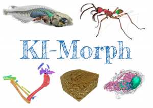
KI-Morph – Petabyte-scale image analysis

Aims & Objectives
The current rapid development of imaging methods for X-ray tomography has led to an increasing field of applications from all areas of life sciences. Here X-ray tomography is used to create 3D data of tissues, down to individual cells and their compartments and up to tissue groups and organs to whole organisms. This 3D data is subsequently used to create 3D models and analyze the morphology of the involved specimen.
While the generation of such 3D data using tomographs is mostly automatic and only takes a few minutes, the image processing and analysis is still mainly manual labor, costing even experienced researchers several orders of magnitude more time. Accordingly, the image analysis step is the major bottleneck in the entire processing pipeline. In our novel research project, KI-Morph, we aim to address this challenge by providing a framework for petabyte-scale image processing and analysis.
Research topics
We are focusing on three main parts:
- Providing the infrastructure for the processing of petabyte-scale imaging data
- Development of AI-based algorithms for the segmentation of large-scale 3D tomography data
- Evaluation of the processing pipeline with data from many areas of life sciences
Funding
The project is funded by the Federal Ministry of Education and Research (BMBF), 2023-2026.
People
Partners
- Engineering Mathematics and Computing Lab (EMCL), Prof. Dr. Vincent Heuveline, IWR, Heidelberg University
- Laboratorium für Applikationen der Synchrotronstrahlung (LAS), Prof. Dr. Tilo Baumbach, Karlsruhe Institute of Technology
- Service-Bereich Future IT – Research and Education (FIRE), Dr. Martin Baumann, University Computing Centre, Heidelberg University
- Centre for Organismal Studies (COS), Prof. Dr. Joachim Wittbrodt, Heidelberg University
- Competence center for biodiversity and integrative taxonomy (KomBioTa), Prof. Dr. Lars Krogmann, Museum of Natural History Stuttgart